This article is part of a broad series on recent advances in the science and medicine of longevity and aging. The series covers a range of topics, including musculoskeletal health. Expect more articles on bone and muscle regeneration to follow.
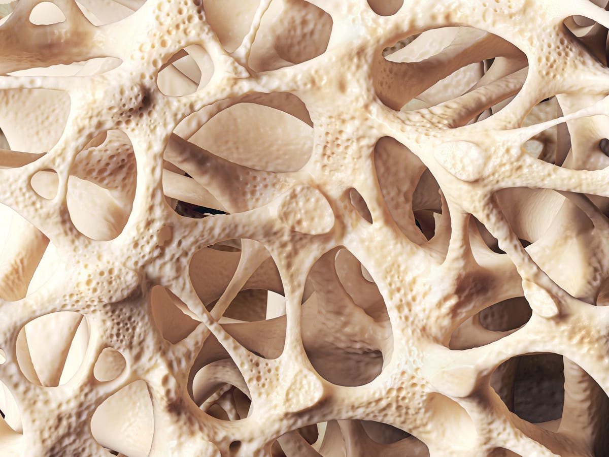
Our weight rests on our bones; they provide structure to everything else. But with age, bone marrow becomes less dense and more “fatty”. The result? Brittle bones that are prone to fracture and struggle to repair themselves. Yet, what happens at a cellular level to cause this deterioration is poorly understood. Researchers at NYU Grossman School of Medicine have now shed light on the underlying mechanisms. Published in Bone Research, an offshoot of Nature, their work uncovers how a signaling pathway that usually helps repair and maintain bone goes haywire as we age, weakening our bones.
Stem Cells and Bone Repair
Bones are special. They are one of the few tissues in the body that can completely regenerate. No scars, no blemishes — as good as new. The unsung heroes in charge of this feat are called skeletal stem and progenitor cells (SSPCs). They reside in the bone marrow, where they help keep things in balance and can be called upon to replace old or damaged bone cells. As stem cells, they are defined by their “plasticity”; the ability to develop into a number of different cell types. Think of them as blank slates that have yet to be given a specific function or purpose. Stem cells residing in the bone marrow can differentiate into two distinct cell types: osteoblasts, which help secrete the scaffolding for bone formation, or adipocytes, which are fat cells that act as stores of energy within the skeletal niche. Between the two, our bones are kept in shape and ready to carry us through life.
Unfortunately, the delicate balance between these two cell types is interrupted as we age. Stem cells begin to develop a bias, preferentially developing into fat cells over their bone-forming counterparts. This leads to a decline in mineral density within the bone marrow and an increase in fat content. Indeed, adipose cells actively release signals that curb the production and regeneration of bone. We are left with “imbalanced” bones that are significantly more fragile and vulnerable to harm.
Notch Signaling: Too Much of a Good Thing?
During early development —while still in the womb— the human body begins to take shape. As part of this process, the skeleton is formed. At first, it’s made up entirely of cartilage. It then slowly turns into bone via ossification. Common to all of these stages is a signaling pathway called Notch, which helps determine cell fate and cell function. The pathway is made up of a family of receptors (Notch1-5) which sit on the surface of cells and facilitate communication between them. Genes involved in this pathway are upregulated during development and help kickstart skeletal formation. Alterations or dysfunction of the pathway, in turn, are associated with a number of skeletal disorders. Once initial development is complete, the signaling pathway is usually downregulated again to allow healthy adult tissue function. And although the Notch pathway continues to play a role in bone health and maintenance throughout life, it is most active during those early developmental stages.
Given its involvement in bone formation and cell fate, Sophie Morgani, Phd, and her colleagues focused on the Notch pathway as a prime suspect for age-related changes to bone marrow. The researchers took bone samples from two, differently aged groups of mice: young-adult mice (3 months old) and middle-aged mice (12 months old). They opted for middle-aged mice instead of aged mice (20 months old) to identify the factors involved in the progression of bone aging and deterioration “rather than in the end product, the irreversibly aged skeleton.”
As expected, the bone marrow of the middle-aged mice had a higher proportion of fat tissue than did the bone marrow of the young-adult mice, suggesting that the skeletal stem cells were skewed towards morphing into adipose cells. Next, the researchers studied the genes being expressed in the bone marrow. Their tests confirmed that key Notch signaling components are upregulated during aging. This was confirmed both by transcriptional and epigenetic tests.
Rescuing Bone Health
If dysregulation of Notch signaling prompts skeletal stem cells to turn into fat cells instead of bone-forming cells, would blocking it help recover bone health?
To test their hunch, the scientists genetically engineered mice to lack nicastrin. Nicastrin is a protein that activates Notch signaling by cleaving all of the Notch receptors; like removing the lid on a bottle, the receptors can suddenly begin to receive and send signals. By blocking nicastrin, you block notch signaling.
Compared to normal middle-aged mice, those that lacked nicastrin had markedly less fat in their bone marrow. On top of this, their bone mass was increased even beyond levels seen in young mice, and their bones’ ability to heal themselves was restored — in effect, the researchers had found a way to fight back against age-related bone degeneration.
The issue is that the Notch pathway is involved in a wide array of different systems, tissues, and cells. Blocking it wholesale would undoubtedly cause unwanted, and dangerous, side effects. Instead, the researchers managed to pinpoint a protein in the signaling pathway that is relatively specific to skeletal stem cells: Early B-cell Factor 3 (Ebf3). This protein is downstream of Notch, so inhibiting it would not have any effect on Notch signaling more broadly. Prior research has noted that the Early B-cell Factor (EBF) family contributes to the build-up of fat deposits in the bone marrow. This makes the protein a promising potential treatment target.
Takeaways
These findings help add an important piece of knowledge to the puzzle of age-related bone deterioration. Although it was known that skeletal stem cells develop a bias towards differentiating into fat cells as we age, why this happens remained unclear. The Notch signaling pathway was the missing link: it becomes dysfunctional with age and contributes to loss of bone density. Blocking the pathway prevents bone degeneration. The work sets the foundation for additional research towards the development of therapeutic strategies.
Read the full article here


